Dilated Pore of Winer
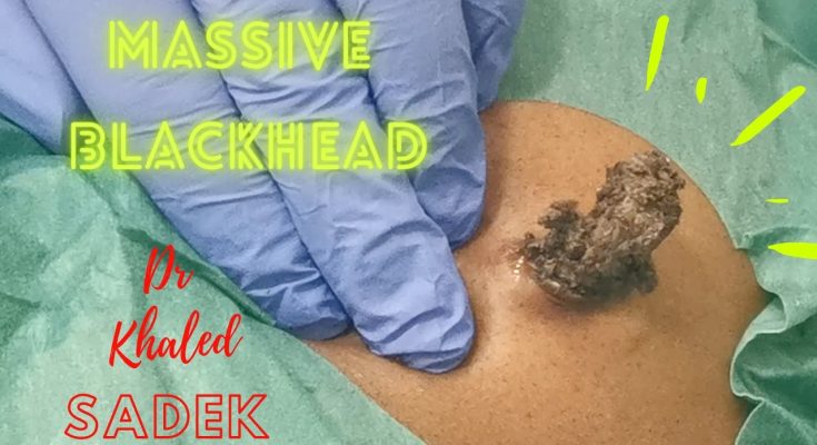
A dilated pore of Winer is a common, giant blackhead pimple, found on your head, neck and torso. Dilated pores of Winer occur on adults and the elderly. Your healthcare provider can easily remove it if you don’t like how it looks on your skin.
What is a dilated pore of Winer?
A dilated pore of Winer is a common, enlarged blackhead pimple (comedo) that originates where hair grows at the hair follicle. A dilated pore of Winer can appear on your head, neck and torso, ranging in size from a few millimeters to more than a centimeter.
What is the difference between a blackhead and a dilated pore of Winer?
The difference between a blackhead and a dilated pore of Winer is size. A dilated pore of Winer is a large blackhead. Both are formed because of clogged pores. A mixture of air and the exposed contents of the clogged pore turn the blemish black (oxidization).
Who does a dilated pore of Winer affect?
A dilated pore of Winer occurs in adults and can appear as early as 20 years old. However, cases usually appear after age 40 and are most common in older ages. Men are more likely to get a dilated pore of Winer, and the tumors are more frequent in people who are white.
Are dilated pores of Winer bad?
Although commonly identified as a tumor, dilated pores of Winer are not a sign of cancer (they are benign). Dilated pores of Winer cannot be spread (non-infectious) and pose no threat to your overall health.
Symptoms and Causes
What are the symptoms of a dilated pore of Winer?
The symptoms of a dilated pore of Winer include:
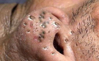
- One enlarged, raised, circular pore covered by a blue to black dot (plug) with normal surrounding skin.
- It is located on the head, neck, face or torso.
- It does not cause any pain or discomfort (asymptomatic).
Does it hurt to have a dilated pore of Winer?
Unless you injure the pore, squeeze or pick it, your dilated pore of Winer should not be painful (asymptomatic). If your dilated pore of Winer is sore, red or leaking pus, it is infected. Make sure you clean the pore as you would a wound and use an antibiotic ointment if you notice an infection. You can also reach out to your healthcare provider to either remove the infected pore or treat it with antibiotics.
What causes a dilated pore of Winer?
A dilated pore of Winer forms similar to a blackhead pimple, where dead skin cells clog the pore (hair follicle). As a result, the dead skin cells in the pore create a protein (sebum and keratin) that collects and plugs up the pore, causing the pore to enlarge (dilate). The clogged pore turns black when the contents of it combine with air exposure (oxidization).
While the direct cause of a dilated pore of Winer is unknown, it has been associated with people who have a history of severe acne and/or cystic acne.
Diagnosis and Tests
How is a dilated pore of Winer diagnosed?
Your healthcare provider can diagnose a dilated pore of Winer by a visual examination. You should not need any test to diagnose this condition.
Management and Treatment
How is a dilated pore of Winer treated?
No treatment is necessary unless your dilated pore of Winer becomes red, swollen and/or leaks pus (infection). You can treat the infection by cleaning the pore and using an antibiotic ointment. If the infection persists, reach out to your healthcare provider to examine the pore and offer treatment options like prescribing an antibiotic to help fight infection.
In most cases, your healthcare provider can remove a dilated pore of Winer if you don’t like how the pore looks on your skin.
How do you fix a dilated pore of Winer?
Smaller dilated pores of Winer can simply be removed with tweezers and a comedone extractor tool that will empty the contents of the pore. When removing the dilated pore of Winer, cleaning out all the contents of the pore reduces the risk of it returning.
If you have a large dilated pore of Winer, don’t try to remove it at home! Your healthcare provider will remove your large, dilated pore of Winer by injecting a small amount of anesthetic near the pore and cutting the skin to remove the contents of the pore. Once the pore is empty, they will stitch the opening of the pore closed. Depending on the size of the pore, stitches are typically removed after 10 days when the wound heals.
How do I close a dilated pore of Winer?
Your healthcare provider will close large dilated pores of Winer with stitches after removing the contents of the pore. Small dilated pores of Winer, similar to the size of a traditional blackhead, should close on their own after squeezing the contents of the pore out with tweezers.
What medications treat a dilated pore of Winer?
Use prescribed antibiotics to treat an infected dilated pore of Winer. No other medications are needed for treatment.
Will my dilated pore of Winer return after removal?
It is likely that a dilated pore of Winer will return after removal if the contents of the pore were not entirely removed. To prevent this, use a skincare routine that cleans pores without clogging them (non-comedogenic).
How long does it take to recover from this treatment?
If your healthcare provider removes the dilated pore of Winer, it could take up to 10 days for the pore to heal.
Prevention
How can I reduce my risk of getting a dilated pore of Winer?
Since the cause is unknown, there is not a method to prevent a dilated pore of Winer from appearing on your skin.
In order to prevent your pores from clogging, you can:
- Use skincare products that don’t clog pores (non-comedogenic).
- Use a cleanser when washing your skin to exfoliate.
- Treat acne when it occurs (use benzoyl peroxide or salicylic acid to dry out dead skin cells).
- Wear sunscreen.
Outlook / Prognosis
What can I expect if I have a dilated pore of Winer?
You can remove a dilated pore of Winer if you don’t like how it looks on your skin, but it isn’t necessary since the pore doesn’t pose any threat to your health.
If your dilated pore of Winer is large, bothersome and you choose to get it removed by your healthcare provider, the likelihood that it will return depends on whether or not they removed the contents of the pore completely. A dilated pore of Winer will return if the pore is still clogged.
Living With
How do I take care of myself?
The best way to take care of yourself if you have a dilated pore of Winer is to avoid touching picking at it or trying to pop the pore it like a pimple. When you bother the pore, it can be painful like a sore or a small wound. Make sure you keep the pore clean and use antibiotic ointment if it becomes infected or irritated.
When should I see my healthcare provider?
If you suspect you have an irritated pore that is red, inflamed and leaking pus or you simply want the pore removed, visit your healthcare provider.
What questions should I ask my doctor?
- Do you recommend I get my dilated pore of Winer removed?
- Will my dilated pore of Winer come back after removal?
- Is my dilated pore of Winer infected?
A note from Cleveland Clinic
While a dilated pore of Winer may look concerning, it isn’t a threat to your health. These large blackhead pimples are common in adults, especially in the elderly, and can be removed if you don’t like how they look on your skin.
Epidermal Inclusion Cyst (Sometimes Called Sebaceous Cyst)
An epidermal inclusion cyst (sebaceous cyst) is a fluid-filled lump under your skin. A keratin substance fills this cyst. It usually doesn’t cause symptoms. Don’t try to pop or remove an epidermal inclusion cyst. A healthcare provider will offer treatment to remove it if it causes discomfort.
What is an epidermal inclusion cyst (sebaceous cyst)?
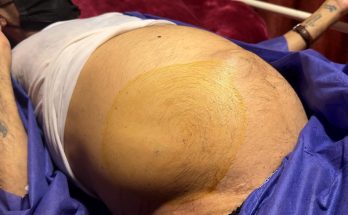
An epidermal inclusion cyst (epidermoid cyst) is a fluid-filled pocket under the surface of your skin. It looks and feels like a lump or bump on your skin.
Many people call epidermal inclusion cysts “sebaceous cysts.” The term “sebaceous cyst” is misleading because the cyst isn’t filled with sebum. Sebum is an oily substance created by your sebaceous glands that keeps your skin moist. Instead, a keratin (protein) and cell debris substance fill epidermal inclusion cysts.
Most healthcare providers only use the term “sebaceous cysts” when associated with the skin condition known as steatocystoma multiplex. Cysts that form with this condition fill with sebum, so they’re truly “sebaceous cysts.” True sebaceous cysts aren’t common, but epidermal inclusion cysts are.
As the name implies, epidermal inclusion cysts form under the top layer of your skin (epidermis).
How common are epidermal inclusion cysts (sebaceous cysts)?
Epidermal inclusion cysts are the most common type of skin cyst.
Symptoms and Causes
What does an epidermal inclusion cyst (sebaceous cyst) look like?
An epidermal inclusion cyst may have the following features:
- A round bump or dome-shaped lump.
- A dark dot (punctum) in the center of the cyst.
- The size ranges from .25 inches to greater than 2 inches. It can grow slowly.
- Skin discoloration (usually pink to red or darker than your natural skin tone).
- Tender or warm to the touch.
- It can move easily.
What are epidermal inclusion cysts (sebaceous cysts) filled with?
A keratin and cell debris substance fills epidermal inclusion cysts. When drained by a dermatologist, this substance looks thick and yellow and has a foul odor.
Is an epidermal inclusion cyst (sebaceous cyst) painful?
An epidermal inclusion cyst isn’t usually painful (asymptomatic). Sometimes, the cyst can inflame (swell) and feel tender when you touch it. As the cyst grows, you may experience skin irritation and pain if it ruptures (breaks open). Occasionally you’ll experience itching at the site of an epidermal inclusion cyst. See your healthcare provider if you develop pain on or near a cyst or have other concerning symptoms.
Where do epidermal inclusion cysts (sebaceous cysts) form?
Epidermal inclusion cysts can form anywhere on your body, but they’re most common on your:
- Face.
- Chest.
- Back.
- Scalp
- Neck.
- Legs.
- Arms.
- Genitalia.
What causes an epidermal inclusion cyst (sebaceous cyst)?
Epidermal inclusion cysts form after a blockage to a hair follicle (an opening in your skin where hair grows out) at the follicular infundibulum (the top part of the hair follicle).
Your body naturally sheds skin cells when they reach the end of their life cycle. If you have a skin injury like a scratch, surgical wound or a skin condition like acne or chronic sun damage, it can disrupt the path your skin cells take to leave your body. This traps these cells and other components like keratin, so they collect under the surface of your skin. This is how a cyst forms.
On areas of your body where you don’t have hair follicles, a cyst can form after an injury or trauma to your skin, too. The injury pushes your skin cells below the top layer of your skin into the second layer (dermis). This creates a pocket where keratin collects and forms a cyst.
What are the risk factors for epidermal inclusion cysts (sebaceous cysts)?
Although they can appear at any age, epidermal inclusion cysts most frequently occur between ages 20 to 60. Epidermal inclusion cysts rarely appear before puberty. They’re more common among people assigned male at birth (AMAB) than people assigned female at birth (AFAB).
Some rare genetic conditions and other conditions lead to the development of multiple epidermal inclusion cysts:
- Gardner syndrome (familial adenomatous polyposis).
- Gorlin syndrome (basal cell nevus syndrome).
- Favre-Racouchot syndrome.
- Human papillomavirus (HPV).
Certain medications may increase your risk of developing epidermal inclusion cysts, including:
- BRAF inhibitors.
- Imiquimod.
- Cyclosporine.
Is an epidermal inclusion cyst (sebaceous cyst) contagious?
No, epidermal inclusion cysts aren’t contagious.
What are the complications of an epidermal inclusion cyst (sebaceous cyst)?
Complications of an epidermal inclusion cysts may include:
- Inflamed epidermal inclusion cyst: The cyst is swollen and tender.
- Infected epidermal inclusion cyst: Your body is fighting harmful bacteria within the cyst, which causes swelling, pain and skin discoloration.
- Ruptured epidermal inclusion cyst: The cyst breaks open, which causes swelling, pain, skin discoloration and yellow (often stinky) fluid drainage.
Is an epidermal inclusion cyst a sign of cancer?
Epidermal inclusion cysts are rarely harmful. However, researchers found rare cases where malignancy (cancer) formed within the cyst, specifically:
An epidermal inclusion cyst may be concerning if it has any of the following characteristics:
- Signs of infection, including pain, skin discoloration, swelling and/or drainage.
- A fast rate of growth.
- A diameter larger than 5 centimeters.
Talk to your healthcare provider if you notice changes to your skin.
Diagnosis and Tests
How is an epidermal inclusion cyst (sebaceous cyst) diagnosed?
A healthcare provider can diagnose an epidermal inclusion cyst during a physical exam simply by looking at it and learning more about your symptoms if you have any.
Although not usually necessary, testing can confirm a diagnosis. It may include:
- Epidermal inclusion cyst radiology or imaging tests: An ultrasound may help determine the contents of the cyst. A CT scan (computed tomography scan) can confirm the diagnosis of a large epidermal inclusion cyst and help your provider determine the best plan for removal.
- A punch biopsy: A provider will remove a small amount of the tissue from the cyst to examine it.
Should I see a specialist for an epidermal inclusion cyst?
If you notice changes to your skin, contact a healthcare provider. You might start with a primary care physician (PCP), and they can refer you to see a dermatologist or a doctor who specializes in skin conditions. Only certain providers can remove epidermal inclusion cysts. Your provider may refer you to a specialist trained to remove cysts, such as a dermatologist, general surgeon or plastic surgeon.
Management and Treatment
How is an epidermal inclusion cyst (sebaceous cyst) treated?
In many cases, a healthcare provider may recommend monitoring the epidermal inclusion cyst and not treating it if it doesn’t cause symptoms.
If the cyst swells and/or causes discomfort, use a warm compress over the cyst to reduce symptoms at home. If your symptoms continue or get worse, contact a provider. They may recommend removing it or they’ll inject a steroid medication into the cyst to temporarily reduce swelling.
Antibiotics can treat an inflamed or infected epidermal inclusion cyst.
Epidermal inclusion cyst (sebaceous cyst) removal
Your provider may remove the epidermal inclusion cyst with the following procedures:
- Incision and drainage: Your provider will make a small opening over the cyst and release the collection of fluid within the cyst. This procedure won’t resolve the cyst since your provider won’t remove the cyst capsule (the outer portion of the cyst). This can help with inflammation and swelling.
- Surgical excision: A surgical procedure that removes the cyst. This procedure uses a local anesthetic (you won’t be asleep and you won’t feel pain). The removal of the capsule (the outer portion of the cyst) prevents the cyst from growing back.
Don’t try popping or draining the cyst yourself. This could cause an infection, and the cyst will likely grow back (recur).
Are there side effects of the treatment?
Risks of surgical excision of a cyst are rare but may include:
- Infection.
- Bleeding.
- Scars.
- Pain.
- Recurrence.
Prevention
Can an epidermal inclusion cyst (sebaceous cyst) be prevented?
Epidermal inclusion cysts typically form randomly. However, avoiding injury or trauma to your skin and treating skin conditions may be helpful to reduce your risk.
Outlook / Prognosis
What’s the outlook for an epidermal inclusion cyst (sebaceous cyst)?
Once you have a diagnosis, you can wait and see if the cyst improves on its own or discuss treatment options with your healthcare provider.
Most cysts don’t cause symptoms. But, it can be challenging if your cyst forms on a very visible part of your body, like on your face or scalp, or if it causes pain. Talk to a healthcare provider about cyst removal if the cyst is bothersome.
Does an epidermal inclusion cyst (sebaceous cyst) go away?
Some cysts decrease in size, while others continue to grow until you get treatment. Without treatment, you may have the cyst for the rest of your life.
Can epidermal inclusion cysts get worse?
Epidermal inclusion cysts sometimes remain small in size and asymptomatic for several years. However, they can also increase in size and may become uncomfortable or irritated. If the cyst bothers you, discuss treatment options with your healthcare provider.
Living With
When should I see a healthcare provider?
Always see your healthcare provider if you find a lump on your skin. It might be an epidermal inclusion cyst, another type of cyst or something else. Don’t try to diagnose it yourself. See your healthcare provider for a clear diagnosis and specialized treatment.
What questions should I ask my healthcare provider?
You may want to ask your provider:
- Do I have an epidermal inclusion cyst or another type of cyst?
- Will this go away on its own, or will it need treatment?
- Do you think the epidermal inclusion cyst will get bigger?
- What treatment options do you recommend?
- Do I need to see a specialist or a surgeon?
- What should I do if the cyst comes back after the procedure?
Additional Common Questions
Is an epidermal inclusion cyst (sebaceous cyst) dangerous?
Most epidermal inclusion cysts aren’t dangerous. They’re usually asymptomatic. Not all epidermal inclusion cysts become infected, but infection is possible. Infections can be dangerous if left untreated. While very rare, some cysts can turn into cancer, so contact a healthcare provider if you notice changes to your skin.
A note from Cleveland Clinic
You may feel scared or anxious after finding a new lump or bump on your skin. The lump may be a harmless epidermal inclusion cyst or it may be a more serious diagnosis. Contact your healthcare provider as soon as you notice changes to your skin. They’ll give you an official diagnosis and answer any questions or concerns you have.
Treatment isn’t always necessary with epidermal inclusion cysts, but you may feel more comfortable if a provider removes it. Don’t try popping or draining the cyst at home. This could lead to an infection. Your healthcare provider will drain the cyst safely, so you don’t have to worry.


