Maggots in Ear: A Rare Case at MAA ENT
Abstract
Myiasis is the infestation of live vertebrates with dipterous larvae. Aural myiasis involves infestation of the external ear and/or middle ear. It is a rare clinical state that usually occurs in patients who have mental or physical disabilities. Although myiasis is a self-limiting disease, it can be associated with fatal complications like penetration within the central nervous system. We present a 87-year-old patient suffering from Alzheimer’s disease with aural myiasis and also discuss the clinical presentation and efective therapies with a review of the literature.
The myiasis, derived from the ancient Greek word ‘myia’ which means fly, is an infestation of the tissues and organs caused by fly (diptera) larvea [1]. Although life cycle of a fly depends on species and types of exposure, usually infestation onsets with flies leaving their ova on intact skin, wound or necrotic tissue. Larvae hatching from the ova pass into adjacent tissues, and complete their life cycles, and transform into adult forms. Myiasis can encounter us in various forms in clinical practice. They are classified as ecologically obligatory, facultative or accidentally localized parasites. Anatomically they are classified according to the location of the larvae in the host [2]. Aural myiasis or automyiasis is the infestation of external ear and/or middle ear with dipterous larvae. This very rarely encountered clinical condition is generally seen in children, in individuals with predisposing factors as mental retardation or impaired personal hygiene. In this article an Alzheimer’s disease patient with automyiasis will be presented after approval of the patient, and their relatives.
CASE REPORT
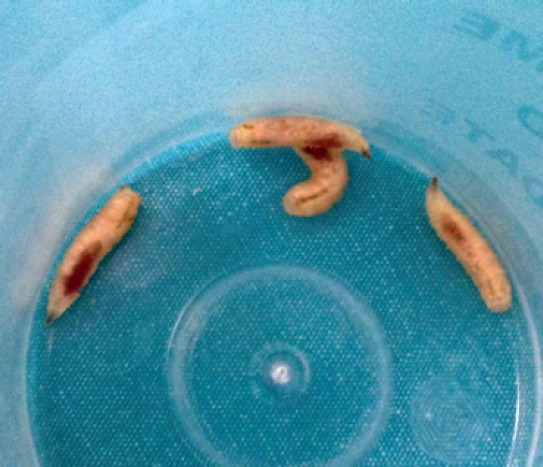
A 87-year-old bedridden woman for 6 months who followed due to severe dementia and monitored in neurology clinic with the diagnosis of Alzheimer’s disease, was consulted to our clinic because of pruritus and bleeding of her left ear. She was suffering from itching in her left ear for nearly a month and bleeding for 2 days. On her physical examination, left external ear canal was completely occluded with mobile larvae (Figure 1). Then the patient was brought into the operating room for otomicroscopic examination External ear canal was irrigated with 10% lidocaine spray, and 70% ethanol to restrict the movements of larvae. Under microscopic guidance, 7 white-colored, thick, and segmented larvae with a mean length of 10-15 mm were extracted from inside the external ear canal with the aid of an aspirator, and alligator clamps (Figure 2). Secretions, and granulation tissue within the external ear canal were removed. An area of perforation on the posterior quadrant of the tympanic membrane was observed. External ear canal, and middle ear were irrigated with physiologic saline. Larvae were not encountered in the middle ear. Topical antibiotherapy was initiated with a mixture of hydrogen peroxide and boric acid solution One week later larvae were not encountered within her external ear canal. Because of her advanced age, and mental health state tympanoplasty operation was not conceived.
FIGURE 1.
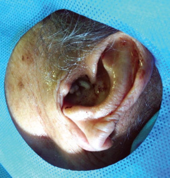
FIGURE 2.
DISCUSSION
Myiasis can be seen in various regions, skin, body cavities, and organs. Aural myiasis can manifest itself in various forms including ophthalmomyiasis, nasal myiasis, urogtenital myiasis, intestinal myiasis, and cutaneous myiasis [3–6]. Only one species of dipterous larva can involve more than one anatomical region or different species can be seen in the same region [2].
Otomyiasis is quiet rarely seen in healthy people [7]. Literature reviews have detected its presence generally in children, people with poor hygiene or those having predisposing factors. Among these predisposing factors mental retardation, immobilization, immunosuppression, and diabetes mellitus can be enumerated [8]. Besides chronic otorrhea is considered as a risk factor in healthy, and ambulatory patients [7]. Our case was a bedridden patient for nearly 6 months with a limited interaction with her environment.
Complaints of otomyiasis can manifest differences based on the patient’s mental health state. These patients can present with complaints of sensation of foreign substance in the ear, aural itching, pain, bleeding, tinnitus, hearing loss, and vertigo [8, 9]. In our case, the most important symptom was itching in the ear. However aural pruritus was not considered as an important symptom by her relatives, and the patient was not consulted to an ENT specialist till her left ear bled. This otorrhagic symptom was thought to be related to frequent scratchings, and the patient was fastened to her bed. Since in cases with otomyiasis early diagnosis is thought to be very important, especially in patients with predisposing factors, persistent ear itching should lead the physician to be suspicious of otomyiasis, and the patients should be evaluated with meticulous otoscopic examination.
In literature reviews, the most frequently encountered species of parasites in cases with aural myiasis are cochliomyia hominivorax, wohlfahrtia magnifica, chrysomya bezziana, chrysomya megacephala, and parasarcophaga crassipalpis [2]. In our case segmented larvae measuring approximately 10-15 mm covered with bands of irregularly, and retrogradely arrayed spinous processes. With these characteristics, these larvae were determined to be consistent with wohlfahrtia magnifica species.
Otomyiasis is generally a self limiting disease. Larvae usually leave the host when they become adult larvae. However during this period because of both mechanical effects of larvae, and collagenases they secrete, they induce many complications in the patient. These complications can include perforation of the tympanic membrane, destruction of the middle ear, and mastoid cavity, and fatal central nervous system invasion [10]. Mortality rates of otomyiasis combined with nasal myiasis climb to 8 percent [2]. The most important point in the prevention of complications is early diagnosis, and eradication of the larvae as soon as possible. The treatment of otomyiasis is basically mechanical cleaning of the airway. This procedure should be performed under microscope is especially important in the evaluation of the middle ear in cases with tympanic membrane perforation. Following mechanical cleaning irrigation of the ear canal with 70% ethanol, 10% chloroform or physiologic saline, urea, dextrose, creatinine, and topical ivermectin is recommended so as to get rid of the remaining larvae. In addition, in cases with suspect furuncle or secondary infection, topical antibiotherapy can be prescribed [8–10].
In conclusion, persistent ear itching, and pain, and irritability especially in patients with predisposing factors as mental retardation, dementia, and immunosuppression, oto myiasis should be kept in mind. A simple autoscopic examination may be a life-saving procedure.
Footnotes
Conflict of Interest: No conflict of interest was declared by the authors.
Financial Disclosure: The authors declared that this study has received no financial support.
REFERENCES
- 1.Hope FW. On insects and their larvae occasionally found in the human body. Trans. R. Entomol. Soc. Lond. 1840:256–71. [Google Scholar]
- 2.Francesconi F, Lupi O. Myiasis. Clin Microbiol Rev. 2012;25:79–105. doi: 10.1128/CMR.00010-11. [DOI] [PMC free article] [PubMed] [Google Scholar]
- 3.Arora S, Sharma JK, Pippal SK, Sethi Y, Yadav A. Clinical etiology of myiasis in ENT: a reterograde period–interval study. Braz J Otorhinolaryngol. 2009;75:356–61. doi: 10.1016/S1808-8694(15)30651-0. [DOI] [PMC free article] [PubMed] [Google Scholar]
- 4.Eyigör H, Dost T, Dayanir V, Başak S, Eren H. A case of naso-ophthalmic myiasis. Kulak Burun Bogaz Ihtis Derg. 2008;18:371–3. [PubMed] [Google Scholar]
- 5.Aydin E, Uysal S, Akkuzu B, Can F. Nasal myiasis by fruit fly larvae: a case report. Eur Arch Otorhinolaryngol. 2006;263:1142–3. doi: 10.1007/s00405-006-0112-0. [DOI] [PubMed] [Google Scholar]
- 6.Karabiber H, Oguzkurt DG, Dogan DG, Aktas M, Selimoglu MA. An unusual cause of rectal bleeding: intestinal myiasis. J Pediatr Gastroenterol Nutr. 2010;51:530–1. doi: 10.1097/MPG.0b013e3181eb313c. [DOI] [PubMed] [Google Scholar]
- 7.Hatten K, Gulleth Y, Meyer T, Eisenman DJ. Myiasis of the external and middle ear. Ann Otol Rhinol Laryngol. 2010;119:436–8. doi: 10.1177/000348941011900702. [DOI] [PubMed] [Google Scholar]
- 8.Güler S, Sağıt M, Sarı K, Somdaş MA. An Aural Myiasis on the Grounds of Chronic Otitis Media: Case Report. KBB ve BBC Dergisi. 2011;19:172–5. [Google Scholar]
- 9.Karaman E, Samasti M, Saritzali G, Ozdemir S, Halil MC, Isildak H. Otomyiasis by Wohlfahrtia magnifica. J Craniofac Surg. 2009;20:2123–4. doi: 10.1097/SCS.0b013e3181bec66e. [DOI] [PubMed] [Google Scholar]
- 10.Werminghaus P, Hoffmann TK, Mehlhorn H, Bas M. Aural myiasis in a patient with Alzheimer’s disease. Eur Arch Otorhinolaryngol. 2008;265:851–3. doi: 10.1007/s00405-007-0535-2. [DOI] [PubMed] [Google Scholar]
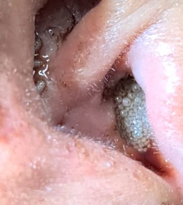
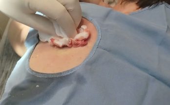
In an unusual and alarming case at Maa ENT Hospitals, an 18-year-old patient presented with an ear infection characterized by foul-smelling discharge due to cholesteatoma. This severe condition inadvertently attracted houseflies, which laid eggs in the ear canal, leading to the development of maggots. Despite the rarity of such an occurrence, the skilled medical team successfully removed the maggots and repaired the patient’s ear.

Understanding the Condition:
Cholesteatoma:
Cholesteatoma is an abnormal skin growth in the middle ear behind the eardrum. It often develops as a cyst or sac that sheds layers of old skin, which can build up and potentially become infected. Symptoms include persistent ear infections, discharge with a foul odor, and hearing loss. If left untreated, cholesteatoma can cause serious complications, including the destruction of ear structures and the spread of infection to nearby areas.
How Maggots Enter the Ear:
Maggots in the ear, or myiasis, occur when fly larvae infest and feed on the necrotic or infected tissues. This condition is particularly rare in modern clinical settings but can happen in cases where wounds or infections are not adequately managed. The flies are attracted to the smell of infection, where they lay eggs that hatch into larvae (maggots), further complicating the infection.
The Case at MAA ENT Hospitals
In this specific case, the patient had been suffering from cholesteatoma, leading to chronic infection and malodorous discharge. The poor hygiene and severity of the infection created an ideal environment for flies to lay their eggs. The maggots that emerged exacerbated the patient’s condition, requiring immediate medical intervention.
Treatment and Management:
The medical team at Maa ENT Hospitals acted swiftly to address this rare condition. The primary steps included:
Mechanical Removal: Using specialized instruments, the doctors carefully extracted the maggots from the ear canal.
Infection Control: The ear was thoroughly cleaned, and the infected area was treated with appropriate antibiotics to prevent further infection.
Surgical Repair: The underlying cholesteatoma was surgically addressed to prevent recurrence. This involved removing the abnormal growth and reconstructing the ear structures as needed.
Why This Case is Rare
Incidents like this are uncommon due to advancements in medical hygiene and the availability of antibiotics. However, certain factors can increase the risk:
Poor Hygiene: Neglect in maintaining ear hygiene can lead to severe infections.
Environmental Conditions: Warm and humid environments can promote fly activity.
Delay in Treatment: Prolonged untreated infections can attract flies, leading to myiasis.
Prevention and Care
To avoid such rare but serious conditions, it is crucial to maintain proper ear hygiene and seek prompt medical attention for any ear infections. Here are some steps to consider:
Regular Cleaning: Ensure ears are kept clean and dry.
Timely Medical Intervention: Do not ignore persistent ear pain or discharge.
Protective Measures: Use ear protection in environments prone to fly activity.
Regular Check-ups: Regular ENT check-ups can help catch and treat conditions early.
While the incident at MAA ENT Hospitals was effectively managed, it serves as a stark reminder of the importance of ear health and hygiene. If you or someone you know experiences symptoms of ear infection, seek medical advice promptly to prevent complications. Stay vigilant about ear hygiene and protect yourself from such rare but possible occurrences.
For more information or to schedule an appointment with our ENT specialists, visit MAA ENT Hospitals today. Your health and safety are our top priorities.
Maggots in the ear can develop under various circumstances. Here, Dr. Meghanadh, the renowned ENT Specialist from MAA ENT Hospital, Hyderabad, explains the different scenarios that can lead to this condition.
